


 Foundational characteristics of cancer include proliferation, angiogenesis, migration, evasion of apoptosis, and cellular immortality. Find key markers for these cellular processes and antibodies to detect them.
 The SUMOplot™ Analysis Program predicts and scores sumoylation sites in your protein. SUMOylation is a post-translational modification involved in various cellular processes, such as nuclear-cytosolic transport, transcriptional regulation, apoptosis, protein stability, response to stress, and progression through the cell cycle.
 The Autophagy Receptor Motif Plotter predicts and scores autophagy receptor binding sites in your protein. Identifying proteins connected to this pathway is critical to understanding the role of autophagy in physiological as well as pathological processes such as development, differentiation, neurodegenerative diseases, stress, infection, and cancer.
CNBP Antibody (Center)
Affinity Purified Rabbit Polyclonal Antibody (Pab)
- SPECIFICATION
- CITATIONS
- PROTOCOLS
- BACKGROUND

Application
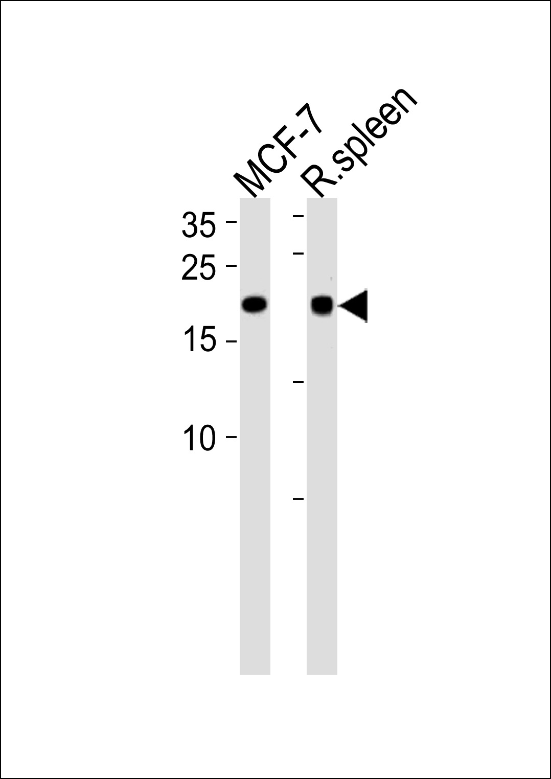
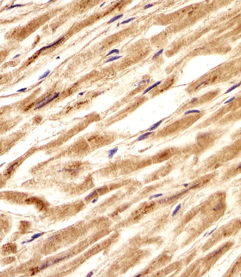
| IHC-P, WB, E |
|---|---|
| Primary Accession | P62633 |
| Other Accession | P62634, P53996, O42395, Q3T0Q6, NP_001120665.1 |
| Reactivity | Human, Rat |
| Predicted | Bovine, Chicken, Mouse |
| Host | Rabbit |
| Clonality | Polyclonal |
| Isotype | Rabbit IgG |
| Calculated MW | 19463 Da |
| Antigen Region | 91-118 aa |
| Gene ID | 7555 |
|---|---|
| Other Names | Cellular nucleic acid-binding protein, CNBP, Zinc finger protein 9, CNBP, RNF163, ZNF9 |
| Target/Specificity | This CNBP antibody is generated from rabbits immunized with a KLH conjugated synthetic peptide between 91-118 amino acids from the Central region of human CNBP. |
| Dilution | IHC-P~~1:25 WB~~1:1000 E~~Use at an assay dependent concentration. |
| Format | Purified polyclonal antibody supplied in PBS with 0.09% (W/V) sodium azide. This antibody is purified through a protein A column, followed by peptide affinity purification. |
| Storage | Maintain refrigerated at 2-8°C for up to 2 weeks. For long term storage store at -20°C in small aliquots to prevent freeze-thaw cycles. |
| Precautions | CNBP Antibody (Center) is for research use only and not for use in diagnostic or therapeutic procedures. |
| Name | CNBP (HGNC:13164) |
|---|---|
| Synonyms | RNF163, ZNF9 |
| Function | Single-stranded DNA-binding protein that preferentially binds to the sterol regulatory element (SRE) sequence 5'-GTGCGGTG-3', and thereby mediates transcriptional repression (PubMed:2562787). Has a role as transactivator of the Myc promoter (By similarity). Binds single-stranded RNA in a sequence-specific manner (By similarity). |
| Cellular Location | Nucleus {ECO:0000250|UniProtKB:P53996}. Cytoplasm. Endoplasmic reticulum {ECO:0000250|UniProtKB:P53996} [Isoform 2]: Cytoplasm [Isoform 5]: Cytoplasm [Isoform 8]: Cytoplasm |
| Tissue Location | Expressed in the liver, kidney, spleen, testis, lung, muscle and adrenal glands. |

Thousands of laboratories across the world have published research that depended on the performance of antibodies from Abcepta to advance their research. Check out links to articles that cite our products in major peer-reviewed journals, organized by research category.
info@abcepta.com, and receive a free "I Love Antibodies" mug.

Provided below are standard protocols that you may find useful for product applications.
Background
This gene encodes a nucleic-acid binding protein with seven zinc-finger domains. The protein has a preference for binding single stranded DNA and RNA. The protein functions in cap-independent translation of ornithine decarboxylase mRNA, and may also function in sterol-mediated transcriptional regulation. A CCTG expansion in the first intron of this gene results in myotonic dystrophy type 2. Multiple transcript variants encoding different isoforms have been found for this gene.
References
Catalli, C., et al. J Mol Diagn 12(5):601-606(2010)
Massa, R., et al. Neuropathol. Appl. Neurobiol. 36(4):275-284(2010)
Sammons, M.A., et al. PLoS ONE 5 (2), E9301 (2010) :
Lucchiari, S., et al. J. Neurol. Sci. 275 (1-2), 159-163 (2008) :
Auvinen, S., et al. Arthritis Rheum. 58(11):3627-3631(2008)
If you have used an Abcepta product and would like to share how it has performed, please click on the "Submit Review" button and provide the requested information. Our staff will examine and post your review and contact you if needed.
If you have any additional inquiries please email technical services at tech@abcepta.com.
Ordering Information
Other Products
Shipping Information
